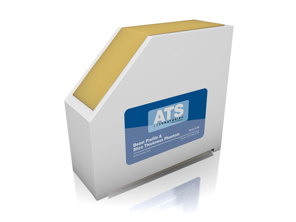
Beam Profile & Slice Thickness Phantom Model ATS 538NH
The CIRS Model ATS 538NH can measure the beam profile and slice thickness of ultrasound imaging systems by evaluating the appearance of a thin plane of echogenic material against an anechoic background.
Overview
Measure Beam Profile and Slice Thickness
The CIRS Model ATS 538NH can measure the beam profile and slice thickness of ultrasound imaging systems by evaluating the appearance of a thin plane of echogenic material against an anechoic background.
Scanning the scattering plane from one surface, perpendicular to the thin plane, obtains an image of the beam profile at varying depths of the Model 538NH. This image contains a great deal of information about the sound beam as it propagates through the tissue-mimicking media such as the focal length, focal zone, beam width, side and grating lobes, and far-field beam divergence. In addition, the near field region of the beam can be easily distinguished from the far field as varying degrees of brightness close to the scan surface versus the homogeneous amplitude further down.
Scanning the scattering plane from a second surface, 45 degrees from the scattering plane, allows users to evaluate the slice thickness of an imaging system at varying depths. Slice thickness or elevational resolution, the third component of spatial resolution, displays reflections produced by structures in front of or behind the beam’s main axis. The effect of changes in the slice thickness is identical to those seen with axial and lateral resolution. The thinner the slice thickness, the better the resolution: as the slice thickness increases, the degree of spatial resolution decreases.
Performance measures:
- Uniformity
- Tissue Harmonic Imaging Compatibility
- Beam Profile/ Focal Zone/ Lateral Response Width